2027 · High School Specialised Subject · Biochemistry
1-4 The Genetic Foundations
DNA, replication accuracy and the path to protein
DNA as the store of information, replication with high fidelity and continuous repair, and the route from a linear sequence to a three-dimensional protein.
3 SECTIONSOutline notesPrint-readyEnglish edition
01
Genetic continuity is based on DNA information storage and transmission
Genes, chromosomes and genomes are three different levels of the same information.
Gene — a segment of DNA whose sequence specifies a functional product (RNA or protein), together with the signals that control its expression
Chromosome — the DNA molecule packaged with proteins; prokaryotes usually carry one circular chromosome, eukaryotes several linear ones
Genome — the complete DNA content of a cell or organism, including coding and non-coding regions
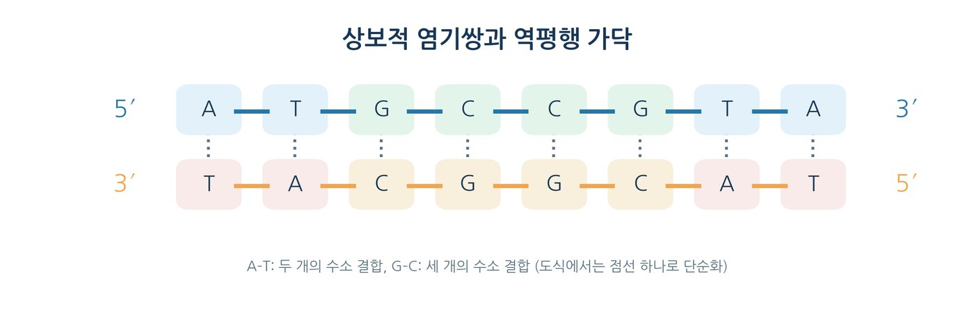
Base pairing — A–T and G–C pairs hold two antiparallel strands together, so one strand templates the other
Antiparallel — one strand runs 5′→3′ while its partner runs 3′→5′
Implication — the sequence is copied with high fidelity and can be repaired, which is what makes heredity stable
DNA is replicated with high accuracy and continuously repaired
Complementary base pairing alone is not accurate enough; proofreading and repair add several orders of magnitude.
Semiconservative replication — each daughter molecule keeps one parental strand and one new strand
Meselson–Stahl — one intermediate band in the first generation, intermediate plus light bands in the second, distinguishes semiconservative from conservative copying
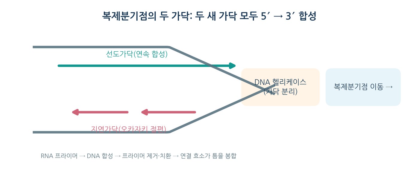
Direction — DNA polymerase adds dNTPs to the 3′-OH end, so the new strand grows 5′→3′ while the template is read 3′→5′
Leading and lagging strands — continuous synthesis on one side, Okazaki fragments on the other
Proofreading — the 3′→5′ exonuclease activity of many replicative polymerases removes misincorporated bases
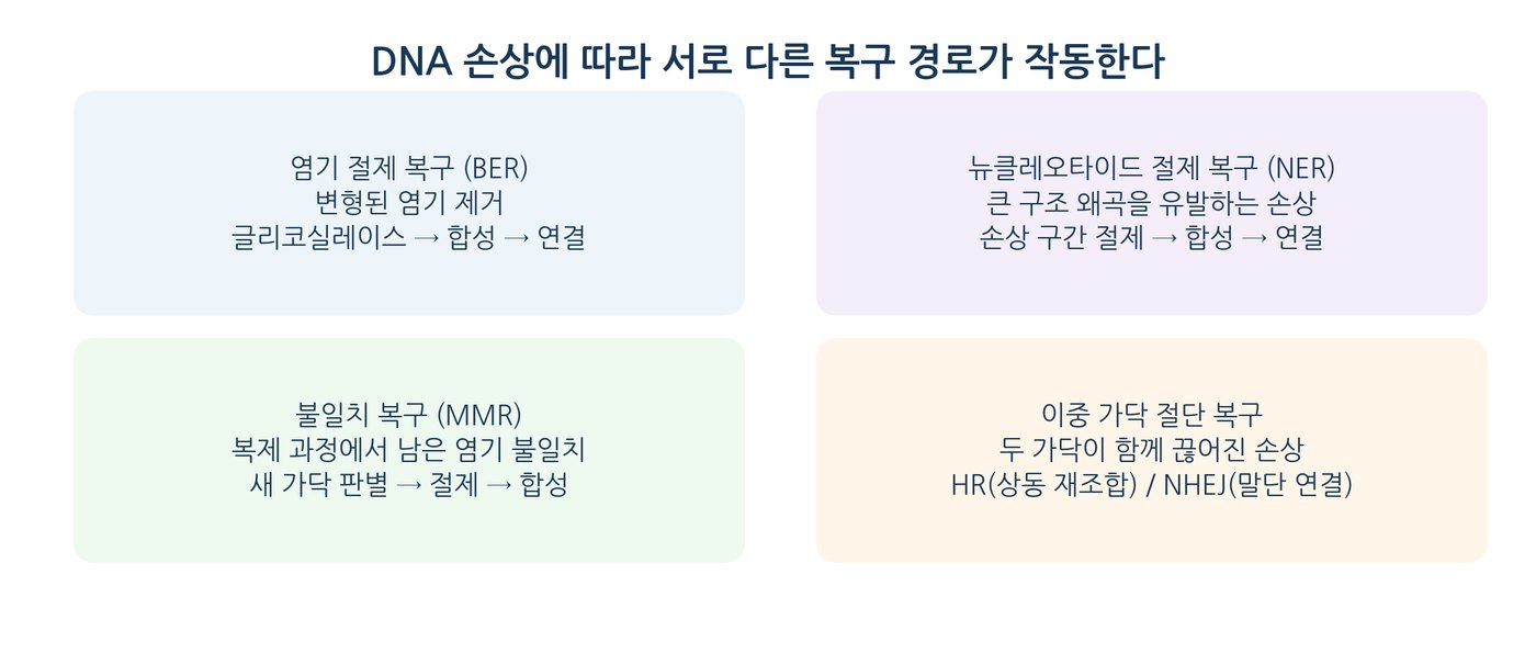
Mismatch repair — corrects errors that survive replication
Error rate — textbooks quote roughly one error per 10⁹–10¹⁰ bases after proofreading and repair; observed mutation rates vary with organism and conditions
Damage ≠ replication error — damage is chemical alteration of DNA; a replication error is a wrong base in the new strand. Unrepaired damage can become a permanent mutation
Repair routes — base excision repair, nucleotide excision repair (UV photoproducts), mismatch repair, homologous recombination and non-homologous end joining for double-strand breaks
Expected errors — E = Np; for N = 4.0 × 10⁶ and p = 1.0 × 10⁻⁹, E = 4.0 × 10⁻³ and P(≥1) ≈ 0.40 %
New strand: 5′ → 3′ · template read: 3′ → 5′
E = Np · P(≥1) ≈ 1 − e−Np
NOTERepair is not always perfect restoration. Depending on the pathway chosen, repair can leave small insertions, deletions or rearrangements.
Noncovalent or some covalent bonds between subunits
Table 1.4-6Model predictions compared with observation
Observation or prediction
Generation 1
Generation 2
Generation 3
HL fraction, semiconservative model
100%
50%
25%
LL fraction, semiconservative model
0%
50%
75%
Feature to compare with the data
One intermediate band
Intermediate plus light band
Rising light-band fraction
Table 1.4-7Expected errors by error probability
Mean error probability p
Expected errors per million bases
Expected errors for N = 2 × 10⁶
10⁻⁵
10
20
10⁻⁷
0.1
0.2
10⁻⁹
0.001
0.002
Table 1.4-8Effect of a substitution and a single-base deletion
Sequence type
mRNA (5′→3′)
Translation product
Reference
AUG GAA UUU UGA
Met–Glu–Phe–Stop
Substitution
AUG GAC UUU UGA
Met–Asp–Phe–Stop
Single-base deletion
AUG AAU UUU GA…
Met–Asn–Phe–…
Table 1.4-9Key terms summarised in one sentence
Term
Summary
Antiparallel and complementary
The two DNA strands run in opposite directions, and A–T and G–C pairing allows template copying
Semiconservative replication
Each daughter DNA molecule has one parental strand and one new strand
Replication accuracy
Base selection, proofreading and mismatch repair greatly reduce errors
DNA damage repair
BER, NER, HR and NHEJ act according to the type of damage
Transcription
RNA is synthesised 5′→3′ on the DNA template
Translation
The ribosome decodes mRNA codons to synthesise a polypeptide
Protein folding
Sequence and environment together determine three-dimensional structure and dynamic function
Table 1.4-10The standard genetic code
First two bases
Third base U
Third base C
Third base A
Third base G
UU
UUU·Phe
UUC·Phe
UUA·Leu
UUG·Leu
UC
UCU·Ser
UCC·Ser
UCA·Ser
UCG·Ser
UA
UAU·Tyr
UAC·Tyr
UAA·Stop
UAG·Stop
UG
UGU·Cys
UGC·Cys
UGA·Stop
UGG·Trp
CU
CUU·Leu
CUC·Leu
CUA·Leu
CUG·Leu
CC
CCU·Pro
CCC·Pro
CCA·Pro
CCG·Pro
CA
CAU·His
CAC·His
CAA·Gln
CAG·Gln
CG
CGU·Arg
CGC·Arg
CGA·Arg
CGG·Arg
AU
AUU·Ile
AUC·Ile
AUA·Ile
AUG·Met
AC
ACU·Thr
ACC·Thr
ACA·Thr
ACG·Thr
AA
AAU·Asn
AAC·Asn
AAA·Lys
AAG·Lys
AG
AGU·Ser
AGC·Ser
AGA·Arg
AGG·Arg
GU
GUU·Val
GUC·Val
GUA·Val
GUG·Val
GC
GCU·Ala
GCC·Ala
GCA·Ala
GCG·Ala
GA
GAU·Asp
GAC·Asp
GAA·Glu
GAG·Glu
GG
GGU·Gly
GGC·Gly
GGA·Gly
GGG·Gly
Figure 1.4-1Genetic variation and its transmission through replication.Figure 1.4-5Three stages that raise the accuracy of DNA replication (illustrative error rates).Figure 1.4-8Substitution and single-base deletion changing the translation output.