01
The cell is the structural and functional unit of life
- Biochemistry — explains, at the molecular level, how biomolecules obeying ordinary chemical and physical laws interact to produce the properties of life
- Six properties of living organisms — chemical complexity and organisation, energy extraction and conversion, precise regulation, sensing and response, self-replication and self-assembly, gradual evolution
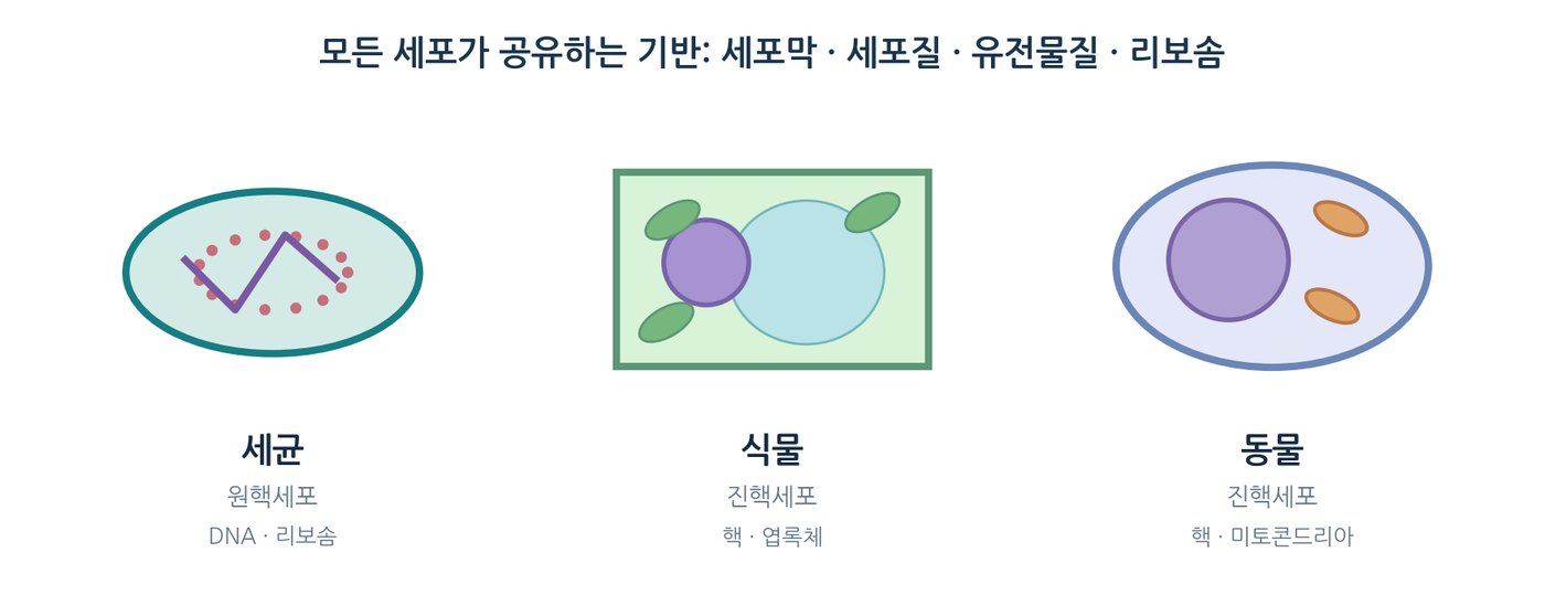
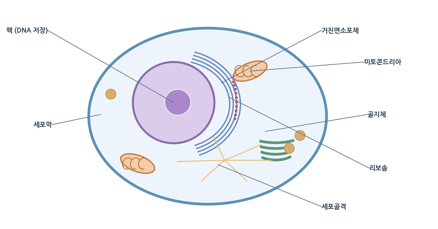
- Plasma membrane — a selectively permeable fluid mosaic of phospholipid bilayer with transport, receptor and signalling proteins; maintains membrane potential
- Cytoplasm ⊃ cytosol — cytoplasm is everything inside the membrane; cytosol is the gel-like, crowded aqueous phase excluding the nucleus or nucleoid, where glycolysis, the pentose phosphate pathway, fatty acid synthesis and protein synthesis occur
- Genome — every cell carries DNA for at least part of its cycle: a membrane-free nucleoid in prokaryotes, a double-membrane nucleus with linear chromosomes in eukaryotes
01
The cell is the structural and functional unit of life
- Biochemistry — explains, at the molecular level, how biomolecules obeying ordinary chemical and physical laws interact to produce the properties of life
- Six properties of living organisms — chemical complexity and organisation, energy extraction and conversion, precise regulation, sensing and response, self-replication and self-assembly, gradual evolution
- Plasma membrane — a selectively permeable fluid mosaic of phospholipid bilayer with transport, receptor and signalling proteins; maintains membrane potential
- Cytoplasm ⊃ cytosol — cytoplasm is everything inside the membrane; cytosol is the gel-like, crowded aqueous phase excluding the nucleus or nucleoid, where glycolysis, the pentose phosphate pathway, fatty acid synthesis and protein synthesis occur
- Genome — every cell carries DNA for at least part of its cycle: a membrane-free nucleoid in prokaryotes, a double-membrane nucleus with linear chromosomes in eukaryotes
02
Cell size is constrained by diffusion
- Lower bound — independent metabolism and reproduction need space for a genome, hundreds of enzymes, ribosomes, tRNA and membrane components; Mycoplasma at 0.1–0.2 μm is close to the theoretical minimum
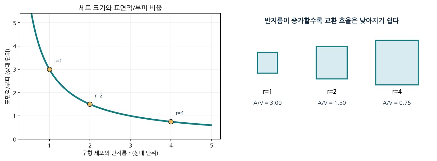
- Upper bound — metabolic demand scales with volume while exchange scales with surface area; most animal and plant cells are 5–100 μm across
- Scaling — S = 4πr², V = (4/3)πr³, so S/V = 3/r; a 10× larger radius gives 100× the surface but 1,000× the volume
- Diffusion time — t = x²/(6D): a 10× longer path costs 100× the time
- Consequence — beyond a certain size the cell core cannot be supplied fast enough and reaches metabolic suffocation
- Four strategies — maximise surface (microvilli), change shape (neurons, muscle fibres), re-partition space (central vacuole), and active transport (cytoplasmic streaming, vesicle traffic)
S/V = 3/r · t = x²/(6D)
02
Cell size is constrained by diffusion
- Lower bound — independent metabolism and reproduction need space for a genome, hundreds of enzymes, ribosomes, tRNA and membrane components; Mycoplasma at 0.1–0.2 μm is close to the theoretical minimum
- Upper bound — metabolic demand scales with volume while exchange scales with surface area; most animal and plant cells are 5–100 μm across
- Scaling — S = 4πr², V = (4/3)πr³, so S/V = 3/r; a 10× larger radius gives 100× the surface but 1,000× the volume
- Diffusion time — t = x²/(6D): a 10× longer path costs 100× the time
- Consequence — beyond a certain size the cell core cannot be supplied fast enough and reaches metabolic suffocation
- Four strategies — maximise surface (microvilli), change shape (neurons, muscle fibres), re-partition space (central vacuole), and active transport (cytoplasmic streaming, vesicle traffic)
S/V = 3/r · t = x²/(6D)
03
Life is divided into three domains
- Old view — prokaryotes versus eukaryotes, judged by the nuclear envelope
- Turning point — Woese and colleagues compared 16S and 18S rRNA sequences in the late 1970s
- Molecular clock — rRNA genes occur in every organism and are highly conserved, making them a reliable measure of evolutionary distance
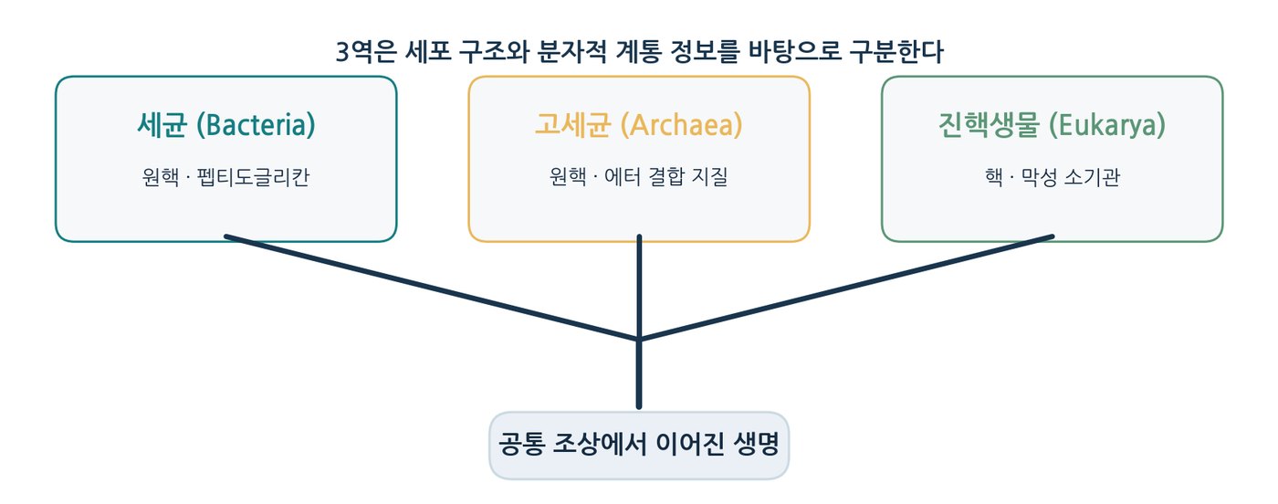
- Three domains — Bacteria, Archaea, Eukarya; the former "prokaryotes" are two lineages that diverged at the base of the tree
- LUCA — all three domains descend from a last universal common ancestor roughly 3.5–3.8 billion years ago; Archaea are closer to Eukarya than to Bacteria
- Dual genetic origin — information-processing genes come from the archaeal line, operational metabolism from the bacterial line
03
Life is divided into three domains
- Old view — prokaryotes versus eukaryotes, judged by the nuclear envelope
- Turning point — Woese and colleagues compared 16S and 18S rRNA sequences in the late 1970s
- Molecular clock — rRNA genes occur in every organism and are highly conserved, making them a reliable measure of evolutionary distance
- Three domains — Bacteria, Archaea, Eukarya; the former "prokaryotes" are two lineages that diverged at the base of the tree
- LUCA — all three domains descend from a last universal common ancestor roughly 3.5–3.8 billion years ago; Archaea are closer to Eukarya than to Bacteria
- Dual genetic origin — information-processing genes come from the archaeal line, operational metabolism from the bacterial line
04
Organisms obtain energy and biosynthetic precursors from different sources
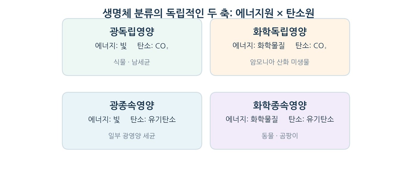
- Energy axis — phototrophs use light; chemotrophs use chemical energy from oxidation–reduction reactions
- Carbon axis — autotrophs reduce CO₂; heterotrophs use preformed organic compounds
- Photoautotrophs — plants, algae, cyanobacteria; oxygenic when water is the electron donor, anoxygenic with H₂S, S⁰ or H₂
- Photoheterotrophs — purple and green nonsulfur bacteria, some halophilic archaea using bacteriorhodopsin
- Chemoautotrophs — sulfur-oxidising, nitrifying and hydrogen bacteria; methanogenic archaea at deep-sea vents
- Chemoheterotrophs — all animals, all fungi, most protists and many bacteria
05
Bacteria and archaea share a plan but differ crucially
- Shared — no nuclear envelope, 0.5–5 μm, a single circular chromosome, 70S ribosomes, plasmids, flagella and pili
- Bacterial wall — peptidoglycan of alternating NAG and NAM cross-linked by peptides; thick in Gram-positive, thin with an outer membrane in Gram-negative
- Difference 1 — no peptidoglycan in archaea: pseudopeptidoglycan or a crystalline S-layer, so penicillin and lysozyme do not act
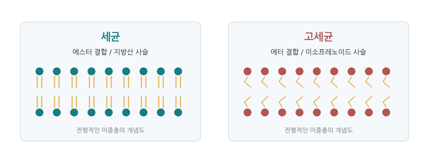
- Difference 2 — ether lipids: D-glycerol, branched isoprenoids and ether linkages instead of ester bonds; GDGT monolayers survive boiling water and strong acid
- Difference 3 — information machinery closer to eukaryotes: RNA polymerase, transcription factors and translation initiation proteins
05
Bacteria and archaea share a plan but differ crucially
- Shared — no nuclear envelope, 0.5–5 μm, a single circular chromosome, 70S ribosomes, plasmids, flagella and pili
- Bacterial wall — peptidoglycan of alternating NAG and NAM cross-linked by peptides; thick in Gram-positive, thin with an outer membrane in Gram-negative
- Difference 1 — no peptidoglycan in archaea: pseudopeptidoglycan or a crystalline S-layer, so penicillin and lysozyme do not act
- Difference 2 — ether lipids: D-glycerol, branched isoprenoids and ether linkages instead of ester bonds; GDGT monolayers survive boiling water and strong acid
- Difference 3 — information machinery closer to eukaryotes: RNA polymerase, transcription factors and translation initiation proteins
06
Membranous organelles separate reactions in space
- Scale — eukaryotic cells are 10–100 μm, roughly 10× the linear dimension and 1,000–10,000× the volume of a prokaryote
- Three benefits — isolate opposing pathways, concentrate enzymes and substrates, and confine hazardous chemistry away from the cytosol
- Nucleus and nucleolus — double membrane with pores; replication and transcription, plus rRNA synthesis and ribosome assembly
- Mitochondria and chloroplasts — double-membrane energy converters; their own circular DNA, 70S ribosomes and division by fission support the endosymbiotic origin
- ER and Golgi — folding, disulfide bonding and N-glycosylation in the rough ER; lipid and steroid synthesis, detoxification and Ca²⁺ storage in the smooth ER; glycan maturation and sorting in the Golgi
- Lysosomes, peroxisomes, vacuoles — acid hydrolases at pH 4.5–5.0; catalase destroying H₂O₂; turgor and storage
07
The cytoskeleton makes structure and transport dynamic
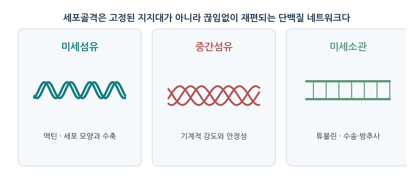
- Functions — mechanical strength, organelle anchoring, chromosome and cytoplasm partitioning, cell movement
- Actin microfilaments — 6–7 nm, ATP-binding G-actin, polar; cortex, microvilli, muscle contraction, contractile ring; motor: myosin
- Microtubules — 25 nm hollow tubes of α/β-tubulin, GTP-binding, dynamic instability; transport tracks, spindle, axoneme; motors: kinesin and dynein
- Intermediate filaments — 8–12 nm, coiled-coil tetramers, no polarity, most stable; resist tension, support the lamina, anchor desmosomes
- Dynamics — treadmilling and dynamic instability reorganise the whole skeleton within seconds to minutes
08
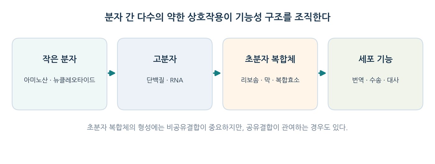
The cell functions as a supramolecular organisation
- Level 1 — monomeric subunits: nucleotides, amino acids, monosaccharides, fatty acids, 100–300 Da
- Level 2 — macromolecules joined by covalent bonds: DNA, RNA, polypeptides, polysaccharides, 10⁴–10⁹ Da
- Level 3 — supramolecular complexes: ribosomes, chromatin, proteasomes, pyruvate dehydrogenase complex, membranes, cytoskeletal fibres
- Level 4 — organelles and the cell, 1–100 μm
- Bonding switch — covalent at 1→2, noncovalent at 2→3: hydrogen bonds, ionic interactions, hydrophobic effect, van der Waals contacts
- Core principle — individually weak bonds acting cooperatively hold complexes together while allowing rapid, low-energy assembly and disassembly
NOTEC–C ≈ 350 kJ/mol · noncovalent 0.4–30 kJ/mol
09
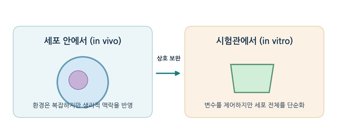
In vitro experiments test interactions between biomolecules
- In vitro — lyse cells, purify the molecule of interest, measure catalysis and binding under controlled temperature, pH and ionic strength
- Limitation — protein at 0.1–1 mg/mL occupies well under 0.1 % of the volume: an extremely dilute world
- In vivo — protein and RNA at 200–300 mg/mL occupy 20–30 % of the cytosolic volume
- Excluded volume — crowding raises effective concentration several-fold above the actual molarity
- Folding and assembly — the drive to minimise excluded volume favours compact folds and multienzyme complexes
- Slower diffusion — large proteins diffuse 5–10× more slowly, favouring substrate channelling along adjacent enzymes
NOTEDilute in vitro solution compared with the crowded in vivo cytosol
10
Key concepts
- Universality — membrane, cytoplasm and genome define a cell
- Size limit — S/V ratio and diffusion time bound cell dimensions; four strategies extend them
- Three domains — Bacteria, Archaea, Eukarya, with eukaryotes closest to the archaeal line
- Four trophic groups — the two axes of energy and carbon source
- Prokaryotic divide — peptidoglycan, ether lipids and information machinery separate bacteria from archaea
- Compartmentation — membranous organelles buy isolation, concentration and protection
- Supramolecular order — weak cooperative bonds give both stability and reversibility
References
| Component | Main chemical composition | Biochemical significance |
|---|---|---|
| Cell membrane | Mainly lipids and membrane proteins | Defines inside and outside; regulates transport and signalling |
| Cytoplasm | Water, ions, metabolites, proteins | The space for metabolism and complex molecular interaction |
| Genetic material | DNA, the cell's hereditary information | Stores the information needed to synthesise enzymes and structural proteins |
| Ribosome | rRNA–protein complex | Reads mRNA and synthesises protein |
| Radius r (μm) | Surface area A (μm²) | Volume V (μm³) | A/V (μm⁻¹) |
|---|---|---|---|
| 1 | ≈12.57 | ≈4.19 | 3.00 |
| 2 | ≈50.27 | ≈33.51 | 1.50 |
| 4 | ≈201.06 | ≈268.08 | 0.75 |
| Domain | Nuclear envelope | Representative organisms | Biochemically important features |
|---|---|---|---|
| Bacteria | Absent | E. coli, cyanobacteria | Cell wall often contains peptidoglycan |
| Archaea | Absent | Methanogenic archaea and others | Membrane lipid chemistry and information machinery differ from bacteria |
| Eukarya | Present | Animals, plants, fungi, protists | Nucleus and membranous organelles divide the labour |
| Feature | Bacteria | Archaea |
|---|---|---|
| Shared cellular features | No nuclear envelope; DNA, ribosomes and a plasma membrane | No nuclear envelope; DNA, ribosomes and a plasma membrane |
| Typical membrane lipid linkage | Fatty acid–glycerol ester bond | Isoprenoid–glycerol ether bond |
| Cell wall | Many species have peptidoglycan | No peptidoglycan; protein layers and other types |
| Transcription machinery | Relatively simple RNA polymerase system | Several components resemble those of eukaryotes |
| Habitat | Very broad | Very broad, including extreme environments |
| Structure | Main function | Biochemical link |
|---|---|---|
| Nucleus | DNA storage and replication, RNA transcription | Genetic information is passed to mRNA |
| Rough ER | Synthesis and early processing of secretory and membrane proteins | Ribosome translation is coupled to protein folding |
| Smooth ER | Lipid synthesis, Ca²⁺ storage, some detoxification | Contributes membrane components and signal control |
| Golgi apparatus | Modification, sorting and transport of protein and lipid | Glycan modification and vesicle traffic |
| Mitochondrion | Aerobic energy metabolism, many intermediates | Inner-membrane electron transport and ATP synthase |
| Lysosome | Breakdown and recycling of macromolecules | Acid hydrolases act at low pH |
| Peroxisome | Oxidation of certain fatty acids, H₂O₂ metabolism | Catalase and related enzymes |
| Chloroplast (plants) | Photosynthesis and carbon fixation | Photochemistry on the thylakoid membrane |
| Element | Main protein and features | Representative functions |
|---|---|---|
| Microfilament | Actin; about 7 nm | Cell movement, cytokinesis, muscle contraction |
| Intermediate filament | Keratin and others; about 10 nm | Mechanical stability; maintenance of nuclear and cell shape |
| Microtubule | α/β-tubulin; outer diameter about 25 nm | Vesicle and organelle transport, cilia and flagella, mitotic spindle |
| Group | Enzyme | Substrate | Purpose |
|---|---|---|---|
| A | Active catalase | Present | Baseline for the normal reaction |
| B | None | Present | No-enzyme control |
| C | Heat-treated catalase | Present | Tests the link between structure and activity |
| D | Active catalase | Absent | Tests whether substrate is required |